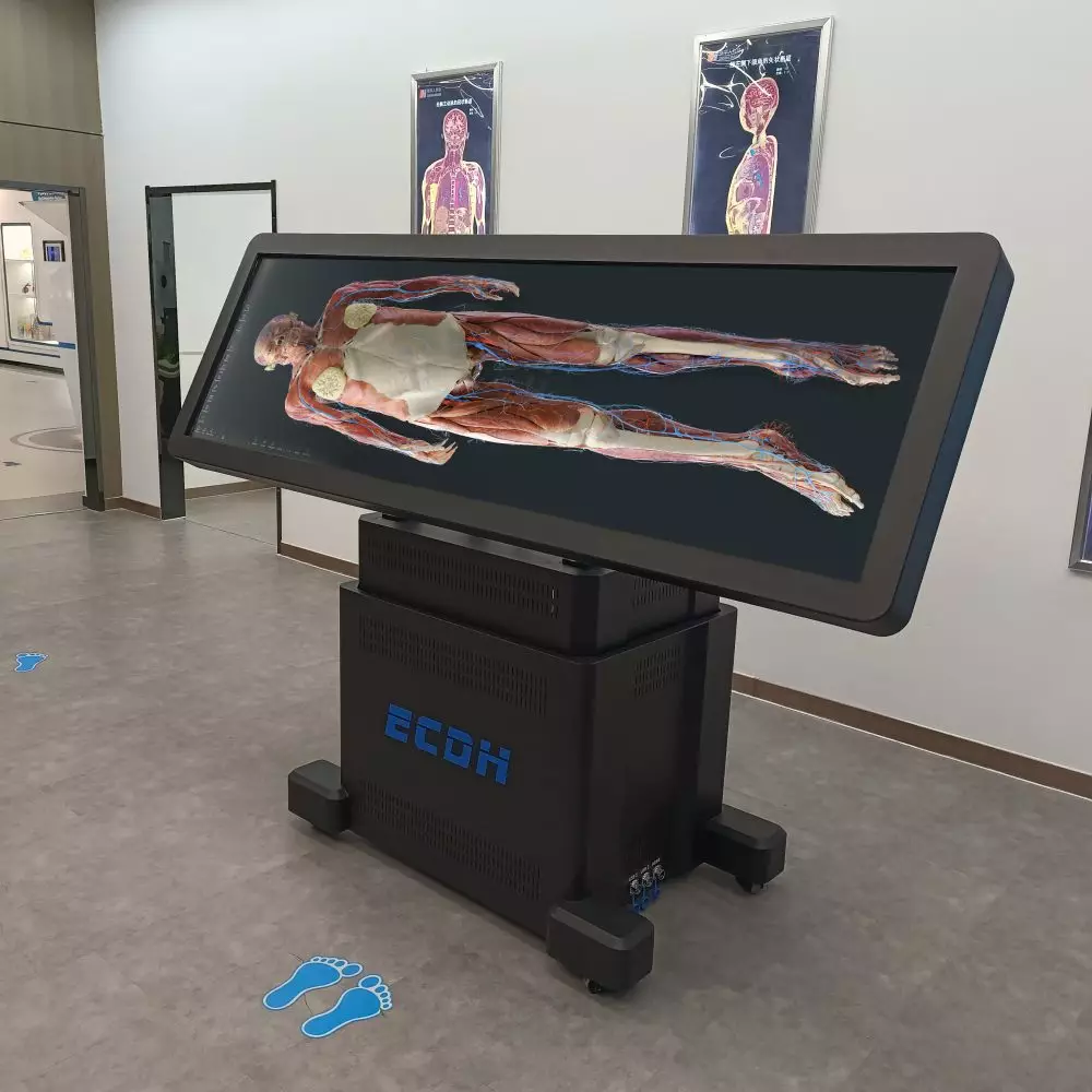
In the modern landscape of medical education, innovative technologies are reshaping how anatomy is taught and understood. The HD Digihuman Virtual Anatomy Table exemplifies this transformation by integrating advanced hardware to enhance the experience of virtual cadaver dissection.
Cutting-Edge Screen Technology for Interactive Learning
At the heart of the HD Digihuman Virtual Anatomy Table is an impressive 88-inch display, offering a resolution of 3840 x 1080 pixels. With a brightness level of 700 cd/m² and a contrast ratio of 1100:1, students can expect crystal-clear images that simulate real anatomy with remarkable accuracy. The touch capability utilizes infrared technology, allowing users to interact with the anatomical models seamlessly during virtual cadaver dissection. Furthermore, the screen’s electric lifting mechanism provides flexibility in teaching scenarios; it can be raised or lowered vertically and tilted up to 90 degrees, accommodating various classroom settings.
Robust Computing Power for Enhanced Performance
The hardware underpinning the HD Digihuman Virtual Anatomy Table is equally impressive. Featuring an Intel I7 processor from the 10th generation, alongside 64GB of DDR4 RAM and a 2TB NVMe SSD, this system is designed to handle complex simulations without lag. The inclusion of an RTX 3080 graphics card ensures that the 3D visualizations are rendered with exceptional detail and fluidity, further enhancing the virtual cadaver dissection experience. Such robust specifications ensure that educators and students can explore intricate anatomical structures interactively and intuitively.
In conclusion, the HD Digihuman Virtual Anatomy Table combines state-of-the-art hardware and innovative technology, revolutionizing the approach to virtual cadaver dissection in medical education. By merging traditional anatomy teaching methods with advanced digital solutions, DIGIHUMAN is paving the way for more effective and engaging learning experiences. This table not only meets but exceeds educational needs, making it an invaluable asset in anatomy laboratories worldwide.


